PATHOLOGY SERVICES WITH HOME COLLECTION AVAILABLE

Signed in as:
filler@godaddy.com
PATHOLOGY SERVICES WITH HOME COLLECTION AVAILABLE

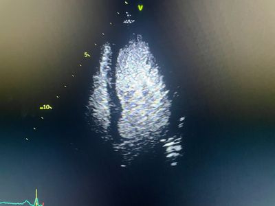
Myocardial Contrast Echocardiography (Contrast Echo) is an advanced echocardiography technique used to improve the visualisation of heart chambers and assess blood flow to the heart muscle more accurately.
A special ultrasound contrast agent made of tiny microbubbles is injected through a vein. These microbubbles enhance the quality of ultrasound images, allowing the cardiologist to detect subtle abnormalities that may be missed on a routine echo.
At HG Superspeciality Clinic, we use contrast echo to improve diagnostic precision in complex cardiac cases.
This is a content preview space you can use to get your audience interested in what you have to say so they can’t wait to learn and read more. Pull out the most interesting detail that appears on the page and write it here.
HG Superspeciality Clinic provides reliable myocardial contrast echocardiography in Thane for accurate assessment of heart function, wall motion abnormalities, structural heart disease, and difficult echocardiographic windows.
A small amount of ultrasound contrast is injected intravenously.
These microbubbles travel through the bloodstream and reflect ultrasound waves more clearly, allowing better visualisation of:
This helps improve diagnostic confidence significantly.
Preparation :
Usually no special preparation is required.
IV Line Placement :
A small intravenous line is inserted for contrast administration.
Contrast Injection :
The ultrasound contrast is injected safely and monitored carefully.
Echocardiographic Imaging :
Real-time images are obtained immediately during contrast circulation.
Duration :
The procedure usually takes 20–30 minutes.